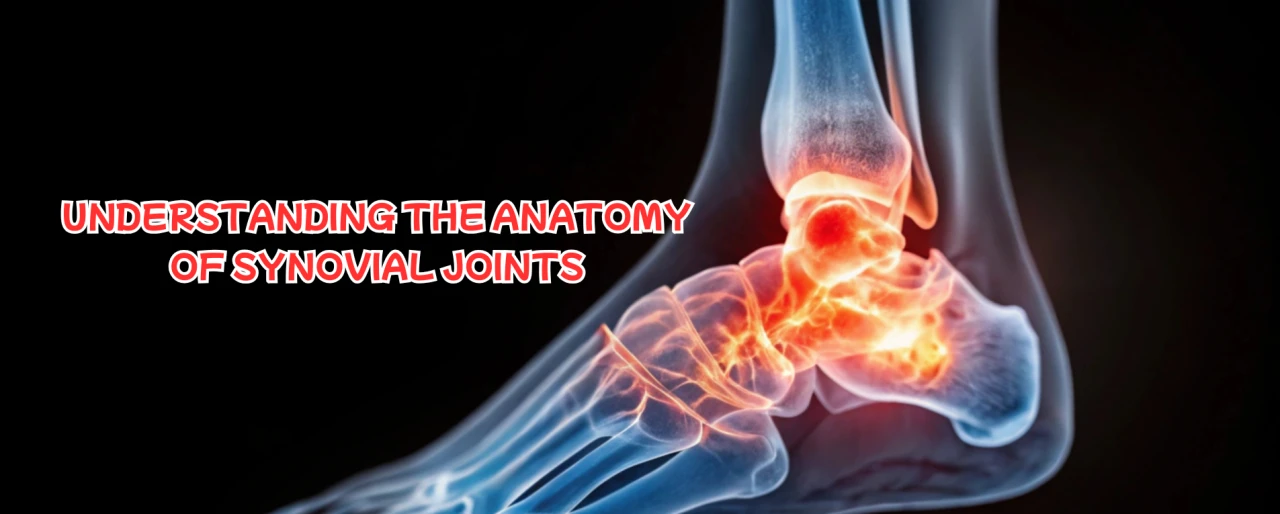
What Are Synovial Joints?
Synovial joints connect two bones through a synovial cavity lined by a synovial membrane, enclosed within a fibrous capsule, and finished with hyaline (articular) cartilage that reduces friction. Classic examples include the knee (tibiofemoral), shoulder (glenohumeral), hip (acetabulofemoral), elbow, wrist, and ankle.
Why it matters: These joints enable walking, lifting, reaching, and grasping. Their high mobility makes them essential to daily function—and vulnerable to sprains, tears, and degenerative disease.
Core Anatomy: Components You Must Know
Articular Cartilage
Hyaline, smooth, avascular surface that minimizes friction and distributes load.
Synovial Membrane & Fluid
Intima/subintima produce hyaluronan-rich fluid for lubrication and cartilage nutrition; viscosity is thixotropic (decreases with shear).
Fibrous Capsule & Ligaments
Capsule blends with periosteum; capsular/extracapsular ligaments guide motion and limit extremes (e.g., ACL/PCL, MCL/LCL).
Menisci, Discs & Labrum
Fibrocartilage structures (knee menisci, TMJ disc, glenoid/acetabular labrum) improve congruency, stability, and shock absorption.
Bursae & Tendon Sheaths
Synovial-lined sacs/tubes that reduce friction between tendons/skin and bone.
Classification: Types of Synovial Joints
| Type | Axes | Examples | Primary Motions |
|---|---|---|---|
| Plane (gliding) | Multi (small) | Intercarpal, intertarsal | Gliding/translation |
| Hinge (ginglymus) | 1 | Elbow, interphalangeal | Flexion–extension |
| Pivot (trochoid) | 1 | Proximal radioulnar, atlanto-axial | Rotation |
| Condyloid (ellipsoid) | 2 | Wrist (radiocarpal), MCP | Flex–ext, abd–add, limited circumduction |
| Saddle (sellar) | 2 | First CMC (thumb) | Flex–ext, abd–add, opposition |
| Ball-and-socket (spheroidal) | 3 | Shoulder, hip | Flex–ext, abd–add, rotation, circumduction |
Biomechanics in Brief
- Stability–mobility trade-off: Deep sockets and strong ligaments (hip) favor stability; shallow sockets with large ROM (shoulder) favor mobility.
- Close-packed vs. loose-packed: Max congruency/ligament tension vs. greatest joint play; relevant for joint mobilization.
- Load distribution: Menisci and cartilage spread compressive forces; damage increases focal stress and degeneration.
- Lubrication: Boundary and fluid-film (hydrodynamic/squeeze-film) mechanisms reduce wear.
Blood Supply & Innervation
Periarticular arterial networks (e.g., genicular vessels at the knee) supply the capsule and synovium. By Hilton’s Law, nerves to muscles moving a joint also supply the joint and overlying skin—explaining referred pain. Capsule and ligaments contain mechanoreceptors essential for proprioception.
Common Pathologies
Osteoarthritis
Cartilage thinning, osteophytes, subchondral sclerosis; pain with load and crepitus.
Rheumatoid Arthritis
Autoimmune synovitis with pannus formation and erosions; morning stiffness improves with activity.
Crystal Arthropathies
Gout/pseudogout inflame synovium; diagnose via crystal analysis after arthrocentesis.
Soft-Tissue Injury
Labral/meniscal tears, sprains/ruptures (e.g., ACL) causing instability, locking, or catching.
Bursitis & Tendinopathy
Overuse friction near high-demand joints (shoulder, knee, ankle) leading to pain and swelling.
Imaging & Assessment
- X-ray: Joint space, osteophytes, alignment.
- MRI: Soft tissue detail—cartilage, labrum, menisci, ligaments.
- Ultrasound: Dynamic assessment of effusions, bursae, tendons; guides injections.
- Arthrocentesis: Diagnostic/therapeutic; fluid analysis (cells, crystals, culture).
Prevention & Care
- Maintain healthy body weight to reduce compressive loads.
- Strengthen periarticular muscles (quadriceps, hip abductors, rotator cuff) for dynamic stability.
- Use neuromuscular training to restore proprioception post-injury.
- Apply ergonomics and graded activity; use proper footwear and technique.
- Seek early evaluation for persistent swelling, instability, or mechanical symptoms.
Also check: 200 Hour Yoga Teacher Training Nepal